The cardiovascular system is a critical topic that is commonly tested on the MCAT, which is why it's so important to include it in your MCAT prep. This system is responsible for circulating blood and delivering oxygen and nutrients to the body's cells, making it essential for maintaining homeostasis and sustaining life. In this article, we will provide an overview of the anatomy and physiology of the cardiovascular system, as well as discuss common cardiovascular diseases and clinical applications.
Disclaimer: MCAT is a registered trademark of AAMC. BeMo and AAMC do not endorse or affiliate with one another.
>>Want us to help you get accepted? Schedule a free initial consultation here <<
Listen to the blog!
Anatomy of the Cardiovascular System
The cardiovascular system is composed of the heart, blood vessels, and blood. Understanding the structure and function of these components is crucial for comprehending how the system functions.
Want to know when to start studying for MCAT? Watch this video:
Heart
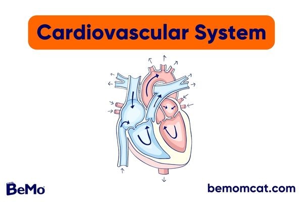
The heart is a muscular organ located in the thoracic cavity. It is responsible for pumping blood throughout the body, providing oxygen and nutrients to the body's tissues and removing waste products. It consists of two upper chambers called atria (singular: atrium) and two lower chambers called ventricles:
- The right atrium receives deoxygenated blood from the body and pumps it to the right ventricle.
- The right ventricle pumps blood to the lungs for oxygenation.
- The left atrium receives oxygenated blood from the lungs.
- The left ventricle pumps oxygenated blood to the body's tissues.
The heart also contains four valves that regulate the flow of blood through its chambers:
- The tricuspid valve is located between the right atrium and the right ventricle.
- The pulmonary valve is located between the right ventricle and the pulmonary artery.
- The mitral valve (or bicuspid valve) is located between the left atrium and left ventricle.
- The aortic valve is located between the left ventricle and the aorta.
The pericardium is a protective sac that envelops the heart to prevent it from overfilling with blood, helping to maintain proper blood flow throughout the body. It consists of two layers:
- The fibrous pericardium provides a sturdy outer layer that helps to anchor the heart in place within the chest and protect it from external trauma.
- The serous pericardium, which is further divided into the parietal layer and the visceral layer, produces a lubricating fluid that reduces friction during the heart's contractions and expansions, ensuring smooth movement.
Blood
Blood is a highly specialized connective tissue that plays a crucial role in the body's overall function. It is composed of various types of cells that are suspended in a liquid called plasma.

Blood coagulation, or clotting, is a critical process in the cardiovascular system that helps to prevent excessive bleeding following injury. The coagulation process involves a complex cascade of reactions that result in the formation of a blood clot.
- Injury to blood vessel wall exposes collagen and tissue factor.
- Platelets adhere to exposed collagen and become activated, releasing clotting factors.
- Clotting factors interact in a cascade, leading to the formation of thrombin.
- Thrombin cleaves fibrinogen into insoluble fibrin strands.
- Fibrin strands aggregate and crosslink, forming a blood clot.
- Anticoagulant factors regulate the clotting process.
Blood Vessels
The circulatory system comprises a complex network of blood vessels that transport blood throughout the body. The table below summarizes the structures and functions of the different types of blood vessels in the cardiovascular system.

The aorta is the largest artery in the body and originates from the left ventricle of the heart and carries oxygenated blood away from the heart to the rest of the body. It has the same three layers as other arteries and branches into smaller arteries, which then further divide into arterioles and capillaries to supply oxygen and nutrients to the body's tissues.
The aorta is divided into three segments:
- The ascending aorta distributes blood to the heart's coronary arteries.
- The aortic arch branches off into major arteries that supply blood to the head, neck, and arms.
- The descending aorta delivers blood to the rest of the body, including the abdomen and legs.
Circulatory System
The circulatory system consists of two distinct circuits that in harmony ensure that oxygen and other vital nutrients are distributed throughout the body and waste products are efficiently removed:
Systemic circulation carries oxygenated blood from the left ventricle to the body's tissues, delivering oxygen and nutrients and removing waste products. Deoxygenated blood is then returned to the right atrium through the veins.
Want to learn more study tips for MCAT Biology? Check out this infographic:
Capillary Beds
Capillary beds are networks of capillaries that allow for the exchange of nutrients, gases, and waste products between the blood and the tissues. They are the smallest blood vessels in the body, with a diameter of about 5-10 micrometers, and are composed of a single layer of endothelial cells. The endothelial cells are connected by tight junctions, which regulate the movement of substances in and out of the capillaries.
There are three types of capillaries:
- Continuous capillaries have a complete endothelial lining, with tight junctions that only allow for the passage of small molecules such as water, ions, and gases.
- Fenestrated capillaries have small pores or fenestrations in their endothelial lining, allowing for the passage of larger molecules such as proteins.
- Sinusoidal capillaries have large fenestrations and discontinuous endothelial lining, allowing for the passage of even larger molecules such as blood cells.
Gas and Solute Exchange
Capillary beds are networks of small blood vessels that allow for gas and solute exchange between the blood and surrounding tissues. The exchange occurs through four main mechanisms:
This is the most common mechanism of gas and solute exchange in capillaries. It occurs because of the concentration gradient of gases and solutes between the blood and the tissues. Oxygen and other small molecules, such as carbon dioxide, can diffuse across the thin walls of the capillaries and enter the surrounding tissues.
Heat Exchange
Heat exchange in capillaries involves the transfer of heat through three mechanisms:
- Conduction refers to the transfer of heat between two objects in direct contact with each other, such as the skin and a cold surface.
- Convection refers to the transfer of heat through the movement of fluids, such as blood circulating through the capillaries.
- Radiation involves the transfer of heat through electromagnetic waves, such as the sun's rays.
Thermoregulation is the process of maintaining a stable body temperature within a narrow range to ensure that metabolic reactions occur efficiently. The cardiovascular system plays a crucial role in thermoregulation by regulating blood flow and heat transfer between the body's core and the skin by adjusting blood flow through vasodilatation/vasoconstriction. This conserves heat and maintains the body's core temperature.
- In warm environments, vasodilation occurs, increasing blood flow to the skin's surface and dissipating heat from the body
- In cold environments, vasoconstriction reduces blood flow to the skin's surface, conserving heat and maintaining the body's core temperature.
The cardiovascular system also plays a role in shivering. As the body shivers, the cardiovascular system works to increase blood flow to the muscles, ensuring they receive the oxygen and nutrients they need to sustain the increased activity. In addition, when the body sweats, blood flow to the sweat glands increases, allowing them to release water onto the skin's surface, where it evaporates and cools the body. The cardiovascular system ensures that blood flow to the sweat glands is adequate to support sweat production and heat loss.
Oxygen Transport by Blood
The cardiovascular system is responsible for the transportation of oxygen from the lungs to the body's tissues and the removal of carbon dioxide from the tissues to be exhaled. This is made possible by hemoglobin, a protein found in red blood cells, which binds to oxygen in the lungs and releases it in the tissues where it is needed for cellular respiration.
- Hematocrit, the percentage of red blood cells in the blood, plays a significant role in oxygen transport as it affects the oxygen-carrying capacity of the blood.
- Oxygen affinity refers to the strength of the bond between hemoglobin and oxygen, which can be affected by factors such as temperature, pH, and the presence of other molecules such as carbon dioxide.
Carbon dioxide produced by cellular respiration is transported in the blood in several forms, including bicarbonate ions, dissolved carbon dioxide, and bound to hemoglobin. The level of carbon dioxide in the blood is regulated by the respiratory system, which controls the rate and depth of breathing to maintain an appropriate balance of carbon dioxide in the blood.
Physiology of the Cardiovascular System
This section explores the intricacies of the cardiovascular system, including the mechanisms behind the cardiac cycle, blood pressure regulation, and blood flow dynamics.
Cardiac Cycle: Systole and Diastole
The cardiac cycle is the rhythmic sequence of events that occur during a single heartbeat, and it consists of two primary phases:
The period during which the heart muscle contracts and pumps blood out of the heart.
However, the cardiac cycle can also be divided into more specific phases based on the activity of individual chambers of the heart. In this case, here is a table summarizing the stages of the cardiac cycle and the corresponding events in the heart:

Blood Pressure
Blood pressure is an essential indicator of cardiovascular health and refers to the force that blood exerts against the walls of blood vessels. Blood pressure is regulated by the nervous system and hormones, such as the renin-angiotensin-aldosterone system (RAAS) and measured using a sphygmomanometer:
- Systolic pressure (the higher number) reflects the maximum force of the heart's contraction
- Diastolic pressure (the lower number) reflects the pressure in the arteries when the heart is at rest.
The body has several mechanisms for regulating blood pressure, including:
Rapid feedback mechanism for maintaining blood pressure within a normal range by decreasing heart rate and vasodilation when blood pressure increases and increasing heart rate and vasoconstriction when blood pressure decreases.
Proper regulation of blood pressure is essential for maintaining a healthy cardiovascular system. Abnormal blood pressure levels can have detrimental effects on the body, leading to cardiovascular disease or organ damage. The ideal blood pressure is typically around 120/80 mmHg, but this may vary based on a person's age, gender, and overall health. Here is a table summarizing the different blood pressure categories based on the American Heart Association's guidelines:

Blood Flow
Blood flow is the volume of blood that flows through a blood vessel in a given period. It is affected by several factors, including blood viscosity, vessel length and radius, and pressure gradient. The relationship between blood flow, pressure, and resistance can be described using Poiseuille's Law equation:
This equation shows that blood flow is directly proportional to the pressure difference and the fourth power of the vessel radius, and inversely proportional to blood viscosity and vessel length. Therefore, changes in vessel radius have a large effect on blood flow.
Cardiac Output
Cardiac output refers to the volume of blood that is pumped by the heart per minute. It is a measure of the effectiveness of the heart in delivering oxygen and nutrients to the body's tissues. The calculation of cardiac output is dependent on two factors: heart rate and stroke volume. Heart rate refers to the number of times the heart beats per minute, while stroke volume refers to the volume of blood pumped by the heart with each beat.
The formula for calculating cardiac output is:
Cardiac Output = Heart Rate x Stroke Volume
For example, if an individual has a heart rate of 70 beats per minute and a stroke volume of 70 milliliters per beat, their cardiac output would be:
Cardiac Output = 70 beats per minute x 70 milliliters per beat
Cardiac Output = 4,900 milliliters per minute or 4.9 liters per minute
The following table lists some of the factors that can increase or decrease cardiac output:

Cardiac Conduction System
The heart is a vital organ responsible for pumping blood throughout the body. To ensure that the heart is functioning properly, it relies on an intricate system of electrical signals known as the cardiac conduction system. This system is made up of specialized cells that generate and transmit electrical impulses through the heart.
The components of the cardiac conduction system include the sinoatrial (SA) node, atrioventricular (AV) node, bundle of His, and Purkinje fibers:
- The SA node serves as the heart's natural pacemaker and generates electrical impulses that cause the heart to beat.
- Electrical impulses from the SA node travel through the atria and reach the AV node.
- The AV node briefly delays the electrical impulses before they continue through the bundle of His and the Purkinje fibers.
- This delay allows the atria to contract and empty blood into the ventricles before the ventricles contract.
Cardiac Action Potential and ECG
Cardiac action potential refers to the electrical changes that occur in the heart muscle cell membrane during the cardiac cycle, leading to the contraction and relaxation of the heart muscle. The action potential is initiated by the flow of ions across the cell membrane, specifically sodium (Na+), potassium (K+), and calcium (Ca2+) ions.
Steps of cardiac action potential:
the resting potential of cardiac cells is around -90mV initiated by the opening of voltage-gated sodium channels, which causes a rapid influx of sodium ions into the cell, and the membrane potential rapidly becomes positive characterized by the entry of calcium ions into the cell, which prolongs the depolarization phase and allows the heart to contract. initiated by the closing of voltage-gated calcium channels and the opening of voltage-gated potassium channels, which causes the cell to become negative again a brief period after repolarization during which the cell cannot be depolarized again, preventing tetanus or summation of action potentials the cell returns to its resting state, with negative membrane potential, and is ready to initiate another action potential
These steps occur during each heartbeat and allow the heart to effectively pump blood throughout the body.
Sample Questions and Answers
In this section, you can find some questions that you can use to test your knowledge! These questions are designed to test your knowledge and understanding of the cardiovascular system.
1. Which type of blood vessel is responsible for exchanging gases and nutrients between the blood and tissues?
A. Arteries
B. Veins
C. Capillaries
D. Lymphatic vessels
2. What is the primary function of the cardiovascular system?
A. To regulate body temperature
B. To transport nutrients and gases to the body's tissues
C. To maintain fluid balance in the body
D. All of the above
3. Which of the following is responsible for carrying oxygen in the blood?
A. Red blood cells
B. White blood cells
C. Platelets
D. Plasma
4. What protein found in red blood cells binds to oxygen in the lungs and releases it in the tissues where it is needed for cellular respiration?
A. Hemoglobin
B. Myoglobin
C. Collagen
D. Keratin
5. Which of the following mechanisms is responsible for gas exchange in capillaries?
A. Active transport
B. Osmosis
C. Diffusion
D. Filtration
6. Which of the following is NOT a type of capillary?
A. Continuous
B. Fenestrated
C. Sinusoidal
D. Atrioventricular
7. What is the term for the resistance encountered by blood as it flows through the blood vessels?
A. Central resistance
B. Systemic resistance
C. Pulmonary resistance
D. Peripheral resistance
8. What is the primary factor affecting peripheral resistance?
A. Blood volume
B. Vessel diameter
C. Vessel length
D. All of the above
9. Which of the following mechanisms is NOT involved in gas and solute exchange in capillaries?
A. Diffusion
B. Facilitated diffusion
C. Osmosis
D. Active transport
10. What is the term for the level of oxygen dissolved in the blood?
A. Hemoglobin saturation
B. Oxygen content
C. Oxygen affinity
D. Hematocrit

