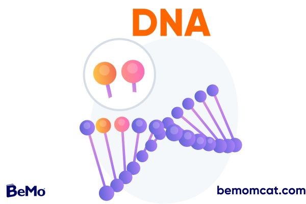
You will certainly be tested on your DNA knowledge on the MCAT. As one of the most fundamental elements of biology, and MCAT biology section, this concept must certainly be present in your MCAT prep. Before you even jump into MCAT practice passages and questions, learn everything you need to know about DNA for your MCAT exam in our article.
Disclaimer: MCAT is a registered trademark of AAMC. BeMo and AAMC do not endorse or affiliate with one another.
>>Want us to help you get accepted? Schedule a free initial consultation here <<
Listen to the blog!
What is DNA?
DNA (Deoxyribonucleic acid) is classified as nucleic acid along with RNA (ribonucleic acid). Nucleic acids are complex biomolecules that serve as the basis for genetic information storage and transfer in living organisms. The structure of DNA as a nucleic acid is essential to its function in carrying genetic information. You may encounter questions about DNA structure, function, replication, and repair on the MCAT. So, understanding DNA as a nucleic acid is crucial for a strong foundation in molecular biology and biochemistry, both important content categories on the MCAT. It will enable you to answer MCAT questions about genetics, molecular biology, and biochemistry. This blog will comprehensively cover the major DNA concepts and its interactions with other biomolecules essential for adequate MCAT preparation.
What are nucleic acids?
The term nucleic acid comes from the Greek word ‘nuclein’ discovered by a Swiss physician Friedrich Miescher, in 1868, from the white blood cells. Nucleic acids are long-chained polymers composed of monomers (nucleotides). These are complex biomolecules crucial for all forms of life. Nucleic acids are found in the nucleus of cells. They transmit genetic material from one generation to the next. Nucleotides are synthesized from readily available cell precursors, such as glucose. DNA and RNA are two types of nucleic acids. DNA controls its replicates, instructs RNA synthesis, and then uses RNA to make protein. This way, genetic information flows from DNA to RNA to proteins called gene expression. The nucleic acids determine each organism’s hereditary traits. However, they are not actively engaged in managing the cell's activities. Instead, the 'proteins' that implement the genetic instruction control how the cell functions.
Nucleotides: nucleic acids’ building blocks
Nucleotides (organic molecules) are the structural units of nucleic acids (DNA and RNA). Each nucleotide comprises a pentose sugar, a phosphate group, and a nitrogenous base. De novo and salvage mechanisms are used for nucleotide synthesis. In de novo synthesis, they are produced from ammonia, carbon dioxide, and biosynthetic components of the metabolism of carbohydrates and amino acids. In the salvage pathway, new nucleotides are created from the nucleotides acquired from the cellular environment and then recovered and recycled. Considering the structure of the nucleotides, each nitrogenous base is composed of one or two rings with nitrogen atoms. They are known as nitrogenous bases because they can absorb H+ from solutions.
Categories of nitrogenous bases
Pyrimidines and purines are the two groups of nitrogenous bases. A pyrimidine consists of a single six-membered ring of carbon and nitrogen elements. Three bases come under the category of pyrimidines:
- thymine (T)
- cytosine (C)
- uracil (U).
In addition, ammonia, beta-amino acids, and carbon dioxide are byproducts of the catabolism of pyrimidines.
Purines consist of two fused rings (six-membered and five-membered rings), so are larger than pyrimidines. There are two purine bases: adenine (A) and guanine (G). Uric acid is produced as a consequence of the breakdown of purines. Both types of nitrogenous bases control the activity of enzymes and are essential for cell signaling. Different chemical groups attached to the rings distinguish the pyrimidines and purines.
Adenine, guanine, and cytosine are found in both DNA and RNA, while thymine is only present in DNA and uracil in RNA. Deoxyribose (DNA) or ribose (RNA) sugars are found in nucleic acids. Both sugars are identical except for the absence of an oxygen atom in deoxyribose on the second carbon in the ring, hence the term deoxyribose.
A condensation reaction is required to bind nucleotides together to form polynucleotides. Phosphodiester linkages link together adjacent nucleotides in the polynucleotide. It comprises a phosphate group that covalently joins the sugars of two nucleotides, creating a sugar-phosphate backbone (repeated pattern of sugar-phosphate units). The polymer's two free ends vary significantly from one another. The polymer's two free ends differ considerably from one another. One end has a phosphate group attached to 5’ carbon and is called a 5’ end; the other is a 3’ end with a hydroxyl group at its 3’ carbon atom. Along the sugar-phosphate backbone, the bases are connected at every point.
Each gene's base sequence on a DNA (or mRNA) polymer is unique and gives the cell a particular kind of information. Genes are hundreds to thousands of nucleotides long, so an infinite number of base patterns could be used. The gene carries the information encodes in its unique arrangement of the four DNA bases. For instance, the sequence 5’-ATGTGCTC-3’ encodes different information than the sequence 5’-CGCAATAC-3’. Bases are arranged linearly in a gene. This sequence specifies the protein's amino acid sequence (primary structure), determining its 3-dimensional shape and role in the cell.
Nucleoside
Nucleoside refers to the part of a nucleotide that contains no phosphate group. So, it consists of a nitrogenous base and a sugar. All living organisms use nucleosides to encode, transmit, and express their genetic information. Through their interactions with purinergic receptors (plasma membrane molecules), nucleosides are crucial for intermediary metabolism, the biosynthesis of macromolecules, and cell communication.
Don't want to study for the MCAT at all? Check out medical schools that do not require the MCAT:
DNA structure: Watson and Crick model
DNA is a double helical structure comprising two strands that spiral around an axis. In 1953, James Watson and Francis Crick first proposed that DNA has a double helix structure. Their model of DNA structure is commonly known as the Watson-Crick model. This concept suggests that the sugar-phosphate backbones are outside each DNA strand, with nucleotides connected by hydrogen bonds inside the helix. The strands are helically twisted, and each right-handed helix comprises ten nucleotides. A helix's diameter is 3.4 nm. The distance between two base pairs that are hydrogen bonded to opposite strands is, therefore, 0.34 nm. The 5' end of one strand faces the 3' end of the other, and the two strands are oriented in opposing orientations, called anti-parallel.
Most DNA molecules have hundreds or even millions of base pairs, which are very long. In addition, DNA contains the nitrogenous bases adenine (A), thymine (T), cytosine (C), and guanine (G). The DNA molecule's specific base sequence forms the genetic code, which controls an organism's traits.
Complementary base pairing
The nitrogenous bases in the Watson-Crick model are always paired in a specific way: adenine bonds with thymine (A-T) and cytosine with guanine (C-G). This complementary base pairing is necessary for DNA replication and the transmission of genetic material from one generation to the next. For example, according to this base pair rule, if one strand has a base sequence of 5’-AGGTCCG-3’, the opposite strand’s sequence must be 3’-TCCAGGC-5’. Due to this feature (complementary pairing), each DNA molecule forms two identical duplicates in a cell, getting ready to divide. The duplicates are shared among the daughter cells during cell division, ensuring they share the parent cell's genetic makeup. Thus, DNA's ability to transmit genetic material during cell reproduction is explained by its structure.
All living things' development, growth, and reproduction depend on DNA's role in the genetic information transmission process. Without DNA, the genetic material necessary for cells, tissues, and organisms to survive and operate would not be passed on from one generation to the next. In addition to storing genetic material, DNA also plays a role in the replication process, mutations, cellular metabolism, transcription, fingerprinting, and gene therapy.
DNA denaturation, reannealing, hybridization
The chemistry of base pairing between complementary strands is responsible for DNA denaturation, reannealing, and hybridization. In DNA denaturation (also known as DNA melting), two strands of DNA are separated. This occurs when the hydrogen bonds between the complementary nucleotides in the two strands are disrupted, usually by raising the temperature or changing the pH of the solution. When DNA is denatured, the individual strands become single-stranded and can no longer base-pair with each other. But this process is reversible.
Single-stranded DNA molecules re-form double-stranded DNA by base-pairing with their corresponding strands, known as DNA reannealing (also called DNA renaturation). This happens when the factors that led to the DNA's denature are removed or altered. For instance, strands reanneal by changing the pH or cooling the solution.
A double-stranded DNA molecule is created during DNA hybridization by binding two complementary single-stranded DNA molecules. This technique is often applied in molecular biology procedures like DNA sequencing and PCR (polymerase chain reaction).
DNA replication
Before cell division, a cell doubles its DNA through DNA replication. This procedure is necessary for cell division and preserving the hereditary material from generation to generation.
Mechanism of replication
DNA replication begins with the unwinding of the double helix. The double helix separates into two single strands when the helicase enzyme breaks the hydrogen bonds linking the two strands. To unzip the DNA helix, the helicase enzyme first catalyzes the creation of a replication fork. It is the point at which two strands split apart. This is essential because high energy intake makes it impossible to unravel DNA strands completely.
Following the separation of the two DNA strands, an enzyme known as DNA polymerase creates new DNA strands by adding nucleotides complementary to each isolated strand. The template is the parental strand, and the freshly synthesizing strands are called daughter strands. The original DNA molecule serves as a template for synthesizing new DNA molecules. The two resulting DNA molecules, known as daughter strands, are identical.
DNA polymerase adds nucleotides to the 3’ end of the daughter strand, and the strand elongates in the 5' to 3' direction. Therefore, the newly synthesized daughter strand is antiparallel to the template strand, which runs in the 3' to 5' direction. As a result, the replication remains continuous when the replication fork moves in a single direction (5' to 3') along the template strand, enabling the polymerase to add nucleotides continuously. The resulting daughter strand, called the leading strand, is a continuous, uninterrupted copy of the template strand.
DNA polymerase enzyme may create a new strand of DNA discontinuously. Discontinuous replication occurs on the lagging strand, which is synthesized opposite from the replication fork's direction. This happens when the replication fork encounters a barrier or an area of the template strand with a complicated structure, causing the DNA polymerase to stop or detach from the template strand. Consequently, many brief DNA fragments known as Okazaki fragments are created in the 5' to 3' direction. Later, a different enzyme known as DNA ligase joins these pieces, creating a continuous, uninterrupted daughter strand.
DNA replication is semiconservative, meaning that each newly synthesized DNA molecule comprises two strands: one original (old) strand and one new strand. Due to this, each daughter molecule includes one original and newly synthesized strand. This has crucial effects on DNA replication because it ensures that every freshly synthesized DNA molecule is a replica of the original DNA molecule. This enables genetic information to be accurately passed from generation to generation.
Enzymes involved in the DNA replication
Several enzymes facilitate DNA replication; each plays a particular role. Together, the following enzymes ensure the precise and effective replication of DNA.
The helicase enzyme unwinds and divides the double helix's two strands at the replication fork so that the DNA polymerase can reach the template strand.
Origin of replication
The origin of replication is the specific position on the DNA helix where DNA replication begins. In prokaryotes, which include bacteria and archaea, the genome is typically circular, and DNA replication initiates at a single origin of replication. For instance, in E. coli, the origin of replication is ‘oriC.’
In eukaryotes, replication has multiple origins because the genome is typically linear. The exact number depends on the size of the genome. Humans have about 30,000 origins distributed throughout their genomes.
Replication of ends of DNA
DNA replication is hampered by linear DNA strands’ telomeres or ends. This is because the function of DNA polymerases prevents the replication machinery from completely replicating the ends. With each round of cell division, the telomeres gradually get shortened. Cells have a specialized enzyme called telomerase to copy the ends of linear chromosomes. This reverse transcriptase uses an RNA template to synthesize DNA at the end of the chromosome. The RNA template base pairs with the telomere, and the telomerase reverse transcriptase uses it as a template to synthesize new telomeric DNA.
DNA repairing
DNA repair is the process cells use to restore damaged or mutated DNA to its original form. Numerous things, such as exposure to radiation, toxins, and errors made during DNA replication, can impair DNA. However, failure to repair DNA damage can lead to mutations and genetic instability, contributing to various diseases, including cancer.
Replication errors can lead to nucleotide mismatches, deletions, or insertions. Cells have developed methods to identify and correct these errors during DNA replication to stop them from accumulating. These mechanisms are collectively known as replication-coupled DNA repair. Proofreading by DNA polymerases is one example of a DNA repair mechanism. Although polymerases are primarily involved in replication, they can also correct errors by proofreading the newly synthesized DNA strand.
Changes in the DNA sequence result in mutations. Some mutations may be neutral or helpful; others may be harmful and cause diseases like cancer. DNA repair mechanisms can fix mutations, such as homologous recombination, non-homologous end joining, mismatch repair, base excision repair, etc. In addition, these mechanisms can detect and repair mutations, such as base substitutions, insertions, deletions, and double-stranded DNA fragments.
DNA mutations: Cancers and other disorders
Mutations in cellular DNA can result in various diseases and tumors. For example, genetic mutations can cause diseases like cystic fibrosis, sickle cell anemia, or Huntington's disease. However, these mutations, which affect every cell in the body, are usually caused by a shift in a single gene.
Two groups of genes that contribute to cancer growth are oncogenes and tumor suppressor genes. When oncogenes are mutated or overexpressed, they cause cancer. Their primary function is to control cell division and development. An oncogene mutation can result in uncontrollable cell division and growth, forming a tumor. Examples include the HER2 gene that produces the HER2 protein. This protein regulates the growth of breast cells. Therefore, a mutation in the HER2 gene is linked to breast cancer. Similarly, overexpression of the BCL2 gene (apoptosis suppressor) is associated with leukemia.
On the other hand, tumor suppressor genes are genes that usually prevent the spread of cancer by controlling cell growth and division, repairing DNA damage, and, when necessary, promoting cell death. When a tumor suppressor gene is mutated, it loses its ability to function, which can cause uncontrolled cell growth and division that eventually results in cancer. For instance, mutated BRAC1 and BRAC2 genes are associated with breast, ovarian, and prostate cancers.

