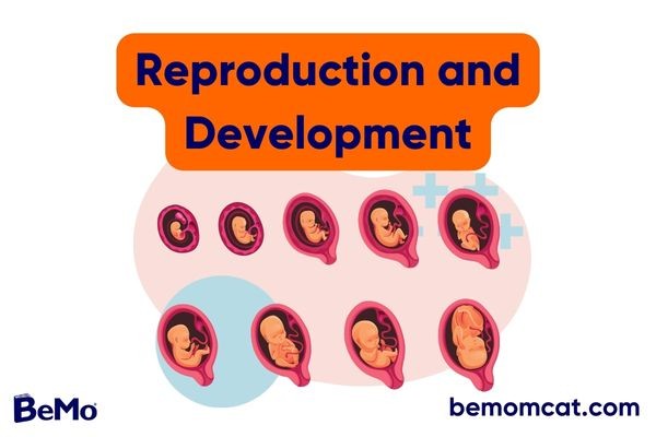
Reproduction and development are fundamental biological processes that involve the fusion of male-female reproductive cells (gametes) and result in offspring that carry genetic material from both parents. Understanding the incredibly complicated processes of reproduction and development is a difficult undertaking that - but it is necessary if you want to get a good MCAT score. Luckily, like with many complex biological concepts, they can be broken down into smaller, simpler ideas!
To help you get started, we have put together a comprehensive guide to exploring the foundational aspects of human reproduction and development and discuss how these concepts are tested on the MCAT. With the help of this guide, you can prepare a solid MCAT study schedule and start your MCAT biology prep right!
>>Want us to help you get accepted? Schedule a free initial consultation here <<
Listen to the blog!
Overview of the Reproductive System
The reproductive system is a collection of tissues, glands, and organs that work together for sexual reproduction. The two main components of the reproductive system are the male reproductive system and the female reproductive system, with the male providing sperm (male gamete) and the female providing the ovum (female gamete).
How to prepare for the MCAT for non-science majors? Watch this video:
Of the body’s major systems, the reproductive system exhibits the most significant differences between sexes and is the only system that does not function until puberty. A network of hormone regulation is essential for the proper functioning of both systems.
Male Reproductive System: Structure and Functions
The male reproductive system performs 3 main functions:
- Produces, maintains, transports, and nourishes sperm and seminal fluid (semen)
- Discharges sperm within the female reproductive tract during sexual intercourse and urine during urination
- Produces and secretes male sex hormones (testosterone)
Most of the male reproductive system exists as external structures located outside of the pelvis, including the penis, scrotum, epididymis, and testes. The testes are the primary reproductive organ (gonads) in males and are responsible for producing sperm and testosterone.
A series of internal organs and ducts, including the vas deferens, seminal vesicles, prostate gland, and bulbourethral glands, aid the process of sperm maturation and transport. During sexual intercourse, chambers in the penis fill up with blood (erection), and sperm is ejaculated in semen by the penis.
Female Reproductive System: Structure and Functions
The female reproductive system consists primarily of internal organs located inside the pelvic cavity, including the ovaries, fallopian tubes, uterus, and vagina. Females also have exterior reproductive structures, such as mons pubis, clitoris, labia majora, labia minora, greater, vestibular glands, and breasts.
Just like the male reproductive system, the female reproductive system also functions to produce gametes and reproductive hormones. However, it also carries the additional task of supporting the developing fetus during pregnancy and delivering it to the outside world.
Here are the 5 main components of the female reproductive system and their functions:
- OVARIES
the gonads that carry/develop ovum and produce hormones (estrogen & progesterone).
- FALLOPIAN TUBES
fertilization sites
- UTERUS
the organ that carries a pregnancy
- CERVIX
regulates sperm entry into the uterus
- VAGINA
the birth canal, which talso enables sexual intercourse
Hormonal Control of Reproduction
Human reproduction and development are largely regulated via reproductive hormones of the hypothalamus, anterior pituitary, and gonads (hypothalamic-pituitary-gonadal or HPG axis).
At the onset of puberty, the hypothalamus secretes high amounts of gonadotropin-releasing hormone (GnRH) to the anterior pituitary gland. In response to GnRH, the anterior pituitary releases two gonadotropins into the blood: follicle-stimulating hormone (FSH) and luteinizing hormone (LH). Both FSH and LH act directly on the gonads of both sexes (testes and ovaries) to regulate gametogenesis.
In males, FSH enters the testes and stimulates the Sertoli cells within the seminiferous tubules to begin spermatogenesis and secrete nourishing factors for sperm cells. LH acts on Leydig cells in the testes and upregulates the production of testosterone, the main male hormone responsible for the stimulation of spermatogenesis and the development of secondary sexual characteristics.
In females, FSH controls ovum production and the maturation of follicles, which are immature cells that assist in the development of the ovum. In. LH, in synergy with FSH, stimulates the developing follicles to produce estrogen and progesterone, the two hormones that regulate the development of secondary sexual characteristics and the onset of the female reproductive (menstrual) cycle composed of three distinct phases:
high FSH levels stimulate the growth of follicles in the ovary. the increased levels of estrogen produced by the follicle result in a surge of LH, leading to ovulation (release of the ovum). high levels of LH continue to promote the formation of the corpus luteum responsible for producing progesterone. The presence or absence of pregnancy determines whether the corpus luteum will continue or cease producing progesterone, either leading to the maintenance of pregnancy or the onset of menstruation, respectively.
Menopause occurs when the ovaries stop producing ovum, as they lose their sensitivity to FSH and LH.
A negative feedback system occurs in the male with rising levels of testosterone and inhibin B (secreted by Sertoli cells) to inhibit the release of GnRH, FSH, and LH and slow down spermatogenesis. In females, negative feedback occurs from high estrogen levels.
Gametogenesis
Gametogenesis refers to the production of sperm in males (spermatogenesis) and ovum in females (oogenesis). This complex process starts with the development of highly specialized germ (precursor) cells located in the testes and ovaries. The result of gametogenesis is the creation of genetically unique gametes to ensure a diverse genetic makeup in the offspring produced through sexual reproduction.
Spermatogenesis
Spermatogenesis is the process through which sperm cells are produced and developed in the male reproductive system. It takes place in the seminiferous tubules in the testes, where the walls are composed of a mixture of developing germ cells and supportive Sertoli and Leydig cells. The least-developed sperm cells are found at the periphery of the tubules, while the fully developed ones are situated in the lumen.
Spermatogenesis can be divided into 4 main stages:
a spermatogonium (2n) divides through mitosis to form a primary spermatocyte (2n), which then undergoes DNA replication to double the amount of genetic material in preparation for meiosis the primary spermatocyte undergo meiosis I to reduce the number of chromosomes from 46 to 23 and form two secondary spermatocytes (n). Both of these secondary spermatocytes then undergo meiosis II, forming a total of four spermatids (n). spermatids develop into functional sperm cells with a flagellum for motility and an acrosome containing enzymes for fertilization. the nearly mature sperm cells enter the epididymis for further maturation until they are ready to be ejaculated during sexual intercourse.
One cycle of spermatogenesis takes 64 days, and a new cycle starts every 16 days, beginning in puberty and continuing throughout a male’s life – although sperm counts may decline after the age of 35.
Oogenesis
Oogenesis is the process of producing the ovum in the outermost layers of the ovaries. While spermatogenesis is initiated only at the time of puberty, oogenesis begins during a female’s development as a fetus and continues until menopause.
The process of oogenesis begins with the mitosis of oogonium (2n) formed during fetal development into the primary oocyte (2n). The primary oocyte will start the meiosis I process and be arrested in its progress during the prophase I stage. Note that all primary oocytes are formed by the fifth month of fetal life and remain dormant in the prophase of meiosis I until puberty.
At each monthly menstrual cycle, one primary oocyte is activated to complete the first meiotic division. The cell divides unequally, with most of the cellular material and organelles going to a secondary oocyte (n) and the rest into a polar body that usually degenerates. During this time, a secondary meiotic arrest occurs, this time at the metaphase II stage.
The secondary oocyte is released during ovulation and travels through the fallopian tube toward the uterus. If the secondary oocyte is fertilized, it continues through meiosis II to form a mature ovum (n), capable of being fertilized by a sperm cell, and a second polar body. If fertilization does not occur, the secondary oocyte is reabsorbed, and the uterine lining is shed, starting the menstrual cycle anew every 28 days.
Want to learn more about MCAT Biology? Check out this infographic:
Fertilization and Early Embryonic Development
The process of human reproduction from a single-celled zygote to a complete multi-cellular organism follows the reproductive sequence of fertilization, implantation, development, and birth. The early stages of embryonic development are crucial for ensuring the overall fitness of the organism.
Fertilization Process
Fertilization is the process by which one sperm cell and one ovum – each carrying a single set (23) of chromosomes – unite to form a zygote. Fertilization occurs in the fallopian tubes, where the ovum is released from the ovary during ovulation. After ejaculation, the sperm cells must swim from the cervix into the uterus, and then up to the fallopian tubes to reach the ovum.
The ovum is protected by a layer of extracellular matrix called the zona pellucida. When a sperm binds to the zona pellucida, a series of biochemical reactions (acrosomal reactions) involving the digestive enzymes present in the acrosome breaks down the matrix for the sperm and ovum membranes fusion. This process creates a pathway for the sperm nucleus to be transferred into the ovum, allowing for the fusion of two haploid (n) genomes to form a new diploid (2n) genome.
Only one sperm cell can fertilize the ovum to ensure that the offspring has only one complete diploid set of chromosomes, and the other sperm cells are typically absorbed into the body. Also, please note that the sperm only contributes half of the DNA, as the ovum actively destroys sperm mitochondria. The ovum contributes another half of the DNA and everything else (mitochondria, organelles, epigenetics).
Cleavage, Blastulation, and Implantation
After fertilization, the zygote begins multiple rounds of rapid mitotic division without increasing in size, a process known as cleavage. The cells then rearrange themselves into a solid ball called morula that later hollows out through a process called blastulation to form a hollow ball called a blastocyst, with a spherical layer of cells (blastoderm) and a fluid-filled cavity (blastocoel).
The blastocyst then continues to divide and begins to differentiate into two layers: an outer shell layer known as the trophoblast and an inner collection of cells called the inner cell mass. The inner cell mass will eventually form the embryonic disk, which will ultimately form the fetus, while the trophectoderm will form the placenta.
Upon reaching the uterus, the embryonic disk attaches itself within the thickened endometrial lining of the uterus through the process called implantation,and pregnancy begins. This attachment is crucial for the transfer of nutrients and oxygen from the mother to the developing embryo.
Gastrulation and Formation of Germ Cell Layers
Gastrulation is a critical stage of embryonic development, in which the blastula transforms into a multilayered structure. The embryonic cells at this stage are extremely pluripotent, meaning they can differentiate into other cell types.
During this time, important signal-calling phenomena (embryonic inductions) set the molecular stage for the initiation of organ formation and three fundamental germ layers that will eventually form all of the tissues and organs of the developing embryo:
- The outermost ectoderm layer gives rise to the nervous system and the epithelial cells.
- The middle mesoderm layer gives rise to different connective tissues and the cardiovascular system.
The innermost endoderm layer gives rise to columnal cells and internal organs that form the digestive system, respiratory system, and urinary system.
Neurulation and Nervous System Development
Neurulation is the process by which the ectoderm differentiates into neurons and supporting neural elements cells to form the neural plate, which will eventually fold and become the neural tube and neural crest. The neural tube will elongate to form the central nervous system (brain and spinal cord). On the other hand, the neural crest will differentiate to form the peripheral nervous system, which includes all the neural cells that extend from the central nervous system to the rest of the body.
Later Embryonic Development
Later embryonic development involves the differentiation of cells into specific tissues and organs, as well as the formation of various structures, such as the heart, nervous system, and digestive system. During this stage, the embryo forms the basic body plan that will eventually lay the foundation for the eventual survival/function of the organism. Any disruptions during this stage can lead to birth defects and other complications.
Stages of Pregnancy
Pregnancy is the state in which a woman carries a developing embryo within her uterus. It typically lasts for about 40 weeks (9 months) from the last menstrual period and is the time during which the developing fetus grows and develops all its organs and systems.
Embryonic development throughout pregnancy can be divided into 3 main stages:
rapid growth and development of the fetus. The neural tube closes, and the heart begins to beat, while the placenta is formed to provide essential nutrients and oxygen to the fetus. the formation of arms, legs, fingers and toes, and the fetus begins to make movements. a time of rapid maturation, with the fetus’ lungs, liver, and brain developing rapidly, and the fetus begins to practice breathing.
Cell Differentiation and Specialization
Cell differentiation and specialization during embryonic development define how generic embryonic cells adopt specific functions and roles. It starts with the commitment of a cell to develop into a specific cell type, followed by its specification and determination.
- Specification is when the cell begins to commit to developing toward a specific cell fate, but the commitment can still be reversed
- Determination is an irreversible commitment of a cell to differentiate based on a specific cell fate.
During cell commitment, the expression of specific genes is turned on/off, leading to the acquisition of specific proteins and other molecular markers that direct the structure, function, and biochemistry of the cells toward specific developmental paths. Cells that have not yet differentiated are known as stem cells.
Cell Communication
Cell communication refers to the process by which cells send and receive intrinsic and/or extrinsic signals that regulate cell behavior and function. This communication can occur between cells of the same type or between different cell types.
There are several different types of cell communication
- DIRECT CONTACT
signals are transmitted directly from one cell to another through cell-to-cell junctions.
- PARACRINE SIGNALING
signaling molecules diffuse from one cell to nearby cells.
- ENDOCRINE SIGNALING
occurs when hormones are produced and released into the bloodstream, traveling to target cells at distant locations.
- SYNAPTIC SIGNALING
occurs between nerve cells and involves the release of neurotransmitters, which bind to receptors on target cells to transmit the signal.
Apoptosis and Regeneration
Apoptosis is a type of programmed cell death that occurs in response to various signals, such as DNA damage or cellular stress. The main purpose of apoptosis is to remove cells that are damaged or no longer needed, thereby preserving the health and function of tissues.
Regeneration, on the other hand, refers to the ability of cells to repair and replace damaged or lost tissues. This can occur through the proliferation of existing cells, the differentiation of stem cells into specialized cells, or the recruitment of new cells from surrounding tissues. These two mechanisms are closely linked, as apoptosis can provide the signals and the cellular building blocks necessary for regeneration to occur.
Sample Questions and Answers
1. What are the female hormones responsible for the regulation of the menstrual cycle?
a. Testosterone
b. Estrogen and progesterone
c. LH and FSH
d. Insulin
2. Which hormone is produced by the corpus luteum and helps to thicken the endometrium?
a. FSH
b. LH
c. Progesterone
d. Estrogen
3. What is the difference between a sperm and an egg cell?
a. Sperm cells are larger and contain more genetic information than egg cells.
b. Egg cells are larger and contain more genetic information than sperm cells.
c. Sperm cells are smaller and contain less genetic information than egg cells.
d. Egg cells are smaller and contain less genetic information than sperm cells.
4. What is the name of the process that occurs in the female reproductive system, in which a fertilized egg implants in the endometrial lining of the uterus?
a. Ovulation
b. Fertilization
c. Implantation
d. Luteinization
5. What is the process by which the zygote divides and becomes a multicellular organism?
a. Fertilization
b. Ovulation
c. Embryonic development
d. Neurulation
6. What is the role of gastrulation in embryonic development?
a. To form the central nervous system
b. To form the peripheral nervous system
c. To differentiate the ectoderm into neurons and supporting neural elements cells
d. To form the three germ cell layers
7. How does the hormonal regulation of the menstrual cycle differ in premenopausal and postmenopausal women?
a. Premenopausal women experience regular fluctuations in hormone levels, while postmenopausal women do not.
b. Postmenopausal women experience regular fluctuations in hormone levels, while premenopausal women do not.
c. Both premenopausal and postmenopausal women experience regular fluctuations in hormone levels.
d. Neither premenopausal nor postmenopausal women experience regular fluctuations in hormone levels.
8. What is the main purpose of apoptosis in embryonic development?
a. To provide signals for cell differentiation
b. To preserve the health and function of tissues
c. To repair and replace damaged or lost tissues
d. To promote the proliferation of existing cells
9. What is the difference between specification and determination in cell differentiation and specialization?
a. Specification can still be reversed, while determination is irreversible.
b. Determination can still be reversed, while specification is irreversible.
c. Both specification and determination are irreversible commitments.
d. There is no difference between specification and determination.
10. Which of the following is not a type of cell communication in embryonic development?
a. Direct contact
b. Paracrine signaling
c. Endocrine signaling
d. Physiological signaling

