Cells and viruses are some of the most high-yield MCAT topics you will encounter during your prep. The MCAT biology section focuses on concepts and understanding of living systems and among its topics, cells and viruses are essential to understand because of a few reasons. First, cells are the basic unit of life, and all living organisms are made up of cells. A thorough understanding of cell structure and function is essential for understanding the human body, including the various organs and systems that make up the body. Second, viruses are important to understand because they can cause a wide range of human diseases. It is crucial for medical professionals to understand how viruses enter cells, replicate, and spread to develop effective treatments and prevent the spread of viral infections. Third, the immune system is vital in defending the body against viral infections. An understanding of how the immune system responds to viral infections is important in the diagnosis and treatment of these infections.
This blog will comprehensively cover the major concepts and topics regarding cells and viruses that are important for effective MCAT prep.
Disclaimer: MCAT is a registered trademark of AAMC. BeMo and AAMC do not endorse or affiliate with one another.
>>Want us to help you get accepted? Schedule a free initial consultation here <<
What is a cell?
The cell is the smallest unit in the body. It is the structural and functional component that sustains life. The number of cells varies in different organisms. There are some single-celled species, like amoebas, and others with trillions of cells, like humans. Cells also differ in size and shape. For example, Mycoplasma gallicepticum is the smallest cell on the earth, and the ostrich egg is the largest. As far as shape is concerned, cells could be spherical (red blood cells), elongated (nerve cells in humans), or spindle-shaped (muscle cells).
Cell discovery and cell theory
Robert Hook first discovered the cell in 1665. Using a microscope, he observed honeycomb-like patterns on a tiny slice of cork. He referred to these entities as cells. Later, more discoveries were made with advancements in the microscope.
German scientists Theodor Schwann and Matthias Jakob Schleiden proposed the cell theory in 1838. The basic principles of the theory are:
- Every living thing is made up of one or multiple cells.
- The cell is the basic unit of life.
- Every new cell comes from an older cell.
Components of the cell
Certain basic features are shared by all cells. These include the plasma membrane, cytosol, chromosomes, ribosomes, and the nucleus. There are also other structures present inside cells performing various essential functions.
Types of cells
Cells are either eukaryotic or prokaryotic based on their structure. The position of DNA is a major difference between eukaryotic and prokaryotic cells. The double membrane-enclosed nucleus of a eukaryotic cell contains the DNA. In comparison, prokaryotic DNA is in a non-membranous structure called a nucleoid.
Prokaryotic and Eukaryotic cells: structure and function
A prokaryotic cell is simpler in structure and function than a eukaryotic cell. It lacks a true nucleus. Other membrane-bound organelles, present in eukaryotic cells, are also absent. Ribosomes are present in the cytoplasm as protein machinery. Prokaryotic cells typically contain only one circular chromosome. They also have plasmids, which are more compact, circular, double-stranded DNA. The cell wall (made up of peptidoglycan), plasma membrane, and glycocalyx (consists of a capsule or slime layer) are external structures of prokaryotes. They have small projections on the surface called fimbriae. These are used as attachment sites. For locomotion, prokaryotes possess one or several flagella.
A prokaryotic cell divides through binary fission. The product of this type of division is two identical daughter cells. The genomic DNA and cytoplasmic contents are distributed equally between the two daughter cells. Examples of prokaryotes include bacteria and archaea.
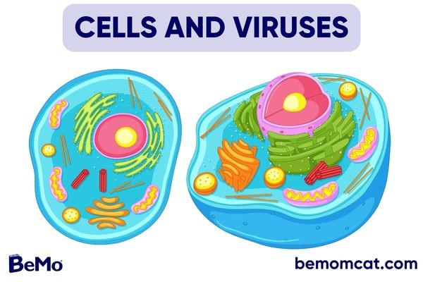
On the other hand, eukaryotic cells possess the true nucleus and complex structure and functions. The plasma membrane is the outermost boundary. It is a fluid mosaic of lipids and proteins. This structure of the membrane maintains its selective permeability. It protects the cell and acts as a gateway for several molecules. It supports cellular transport through endocytosis and exocytosis. In endocytosis, the plasma membrane constricts inward and creates a vesicle, thereby swallowing large molecules. Exocytosis involves the fusion of a vesicle with the plasma membrane, and large molecules are secreted. A rigid cell wall in plant cells surrounds the plasma membrane. It regulates the shape of the plant cell, provides protection, and prevents excessive water absorption. In a eukaryotic cell, a range of organelles is suspended in cytosol. The following specialized structures perform a variety of functions.
Nucleus
The core of the cell contains the nucleus. It processes information, the so-called brain of the cell. A porous, double membrane nuclear envelope separates it from other cellular organelles. Inside the nucleus, chromosomes and nucleolus are present. A chromosome is a thread-like structure of one DNA and many histone proteins. Chromatin is the name given to this DNA and protein complex. The nucleolus is a dense structure. It is a site of synthesis for ribosomal RNA (rRNA). The number of nuclei in a cell depends on its species and stage in the reproductive cycle. In the nucleus, DNA is duplicated, and mRNA is synthesized.
Ribosomes
Ribosomes are called the protein factories of cells. They are composed of RNA and protein. There are two types of ribosomes: free ribosomes and bound ribosomes. Free ribosomes float freely in the cytoplasm, and bound ribosomes are attached to the nuclear envelope or endoplasmic reticulum. Both kinds of ribosomes synthesize different types of proteins. For example, bound ribosomes make membrane proteins or those secreted as enzymes, while free ribosomes synthesize those proteins that remain in the cytosol for various functions, for example, an enzyme used in glycolysis.
Endoplasmic reticulum (ER)
Several membranes form this extensive network in the cytoplasm. They possess cisternae, which are membrane sacs. The inside space of ER is the lumen. The two different types of ER are rough endoplasmic reticulum, which has associated ribosomes, and smooth endoplasmic reticulum. Smooth endoplasmic reticulums are involved in protein synthesis, modification, and transportation. The smooth ER helps various metabolic processes, such as calcium ion storage, glucose metabolism, drug and toxin detoxification, and lipid synthesis.
Golgi apparatus
The Golgi apparatus receives and transports the products of the cell. The group comprises several connected, flattened membrane sacs called cisternae. It processes, stores, and transports proteins and other ER products to their target destinations. Vesicles carry out the transport of material. They are specialized structures for secretion.
Vacuoles
The Golgi apparatus and endoplasmic reticulum produce large vesicles called vacuoles. Animal cells contain many small vacuoles. A single large vacuole provides rigidity in the plant cell. Vacuoles carry out several diverse tasks depending on the cell. For instance, food vacuoles store food, and contractile vacuoles remove excess water from the cell.
Lysosomes and peroxisomes
Lysosomes are called the cell’s suicidal bags. They have hydrolytic enzymes that are used to break down macromolecules. These enzymes work best in an acidic medium. Peroxisomes are specialized metabolic compartments that contain enzymes. These enzymes carry out oxidation reactions and break down hydrogen peroxide.
Mitochondria
Mitochondria are the cell’s powerhouse. Cellular respiration occurs in this organelle that generates adenosine triphosphate (ATP). The organelle is bounded by a double membrane envelope. The inner membrane folds into cristae, while the outer one is smooth. There is an intermembrane space between the two membranes. The inner membrane encloses the mitochondrial matrix. There are several enzymes, mitochondrial DNA, and ribosomes in this matrix.
Plastids
Plastids are found in the cells of both algae and plants. These double-membrane organelles produce and store food. Chloroplasts are a form of plastid that carry out the process of photosynthesis. Flattened sacs called thylakoids are present inside the chloroplasts. They are stacked on top of one another and form granum. A fluid called stroma surrounds the thylakoid. It contains many enzymes, ribosomes, and chloroplast DNA. Another type of plastid is amyloplast. In roots and tubers, it accumulates starch (amylose). Chromoplast plastids have pigments that determine fruits and flowers’ orange and yellow colors.
Cytoskeleton
Cells are supported by a network of fibers called the cytoskeleton. This is vital for animal cells because they don’t have cell walls. These fibers are named microtubules (thick), microfilaments (thin), and intermediate filaments (middle range). Microtubules in animal cells originate from the centrosome. Two centrioles are present within a centrosome. Each has a ring structure made up of nine sets of triplet microtubules.
Flagella and cilia
Flagella and cilia are present in some eukaryotic cells. These are cellular extensions made up of microtubules. These act as locomotor appendages in many unicellular organisms. Flagella are found in the sperm of various plants, algae, and animals. Cilia usually occur on the cell surface and perform multiple functions. For example, mucus in the trachea is swept by its ciliated lining. Cilia are smaller and more abundant than flagella. One or two flagella are typically present in each cell and are longer than cilia.
Not a science major? Check out our tips:
Cell communication
Cells communicate using chemical signals. Signal reception, transduction, and cell response are steps of the signal transduction process. Multicellular and unicellular organisms have various such pathways. Cell communication is either local or over a long distance.
Local signaling
This type of signaling occurs between adjacent cells. Signals that act locally are called paracrine signals. For instance, growth factors in animals are local regulators. They regulate the growth of adjoining target cells. Synaptic signaling is another specific type of local signaling. It takes place in the nervous system of animals. In this process, an electric signal passes along the nerve cell. This signal stimulates the neurosecretory cells in the brain. These cells release neurotransmitter (e.g. acetylcholine), which serves as a chemical signal. The neurotransmitter crosses the synapse (narrow junction between a nerve cell and target cell) and reaches the effectors. The effectors are target cells (neuron or muscle) that respond to the chemical signal.
Long distance signaling
Animals and plants use hormones to extend prolonged distant signaling, called endocrine signaling. Specialized cells release hormones. In animals, these hormones travel through the bloodstream to the target cell, while in plant cells, they travel via other cells. Some common animal hormones are insulin, glucagon, prolactin, oxytocin, thyroxine, etc. Plant hormones include ethylene, auxin, cytokinin, and others.
How do cells divide?
Cells divide through two mechanisms: mitosis and meiosis.
Mitosis
Mitosis is the primary biological process that forms new body cells. Before division, replication of DNA takes place. It is called the interphase of the cell. The G1, S, and G2 phases constitute this phase. The four stages of mitosis are prophase, metaphase, anaphase, and telophase.
The nuclear membrane disappears, chromosomes condense, and the spindle begins to form during the prophase. Two centrioles move toward the opposite poles.
Next comes the metaphase, when chromosomes align at the center of the spindle. With the separation of sister chromatids, the division transitions from metaphase to anaphase. By the end of the anaphase, both chromatids are at opposite poles.
Now the telophase begins, which is the last stage. It is the reverse of prophase; the chromosomes are decondensed, and the nuclear envelope reforms. The telophase is followed by cytokinesis, and the cytoplasm splits into two daughter cells. These cells are genetically similar.
Mitosis is necessary for normal body development and growth. When mitosis is not adequately controlled, health problems like cancer may result.
Meiosis: reduction division
Meiosis is a particular type of division that occurs only in germ cells. Compared to mitosis, meiosis produces four daughter cells. These cells contain haploid chromosomes. Like mitosis, meiosis also proceeds through multiple stages. These phases are similar to mitosis except for a few differences. For example, in mitosis, sister chromatids separate during the anaphase, while in meiosis, homologous chromosomes separate. Meiosis is completed in two rounds: meiosis I and meiosis II.
Four phases follow in each round.
A cell must first pass through the interphase before beginning meiosis I. The diploid germ cell’s chromosomes have all undergone replication. As a result, paired duplicate chromatids have formed.
Prophase I starts, and visible differences appear. Chromosome constriction begins. Unlike mitosis, chromosomes pair up, and tetrad is formed. Each chromosome lines up with its homologous partner. Crossing over takes place between the homologs. In this process, both chromosomes exchange their segments. As a result, recombinant chromosomes are formed. The cell moves into the metaphase, and the spindle captures chromosomes. They are transferred to the cell’s center, called the metaphase plate. Here, the homologous pairs, not individual chromosomes, align for separation. While aligning, the position of each pair is randomly determined. As a result, , the gametes are formed with various sets of homologs. The homologs separate and move to the cell’s opposite ends. This marks the beginning of the anaphase. The telophase starts once the chromosomes have reached their respective poles. The nuclear membrane reforms, and the chromosomes decondense. In the end, two haploid daughter cells are formed via cytokinesis.
What are viruses?
The word “virus” originates in the Latin word meaning “poison.” It is an infectious agent composed of a protein-coated nucleic acid segment (DNA or RNA). Viruses have no independent existence, as they always reproduce inside a host cell. They multiply using the host’s machinery and cellular metabolism. Viruses are the tiniest microbes. Most viruses have a diameter between 20 to 400 nanometers. However, the largest viruses, known as Mimivirus, have diameters greater than 400 nm.
Discovery of viruses
In 1883, the German scientist Adolf Mayer studied a disease called tobacco mosaic. He tried extracting sap from the diseased leaf to search for microorganisms but was unsuccessful. Consequently, he suggested that a bacterium caused the disease. Later, Dmitri Ivanowsky took the sap of an infected tobacco plant and tested it to identify the microorganism. After passing the fluid through a filter paper, he found that the filtrate was able to produce the disease. Then, Martinus Beijerinck conducted a series of experiments. He noticed that the infectious agent in the filtrate could replicate. However, unlike bacteria this infectious agent could not be cultivated on nutrient media. Finally, in 1935, Wendell Stanley, an American scientist, was succeeded in studying the tobacco mosaic virus (TMV). He crystallized the TMV from infected plant leaves and showed that its structure is composed of RNA and protein. So, the era of virus study began.
Here're some more tips for your MCAT prep:
Structure of viruses
The viral genome is enclosed in a protein shell, called a “capsid,” composed of several protein subunits called capsomeres. A virus capsid is helical or icosahedral in shape. The helix is a spiral shape with cylindrical edges that wrap around an axis. For example, the first described virus, TMV, has a naked helical structure.
Both enveloped and uncoated helical viruses exist. The envelope contains viral proteins and the host cell’s phospholipids and membrane proteins. Enveloped helical viruses in plants are uncommon. However, all animal helical viruses, such as influenza, rabies, and mumps, are enveloped.
On the other hand, an icosahedron is a 20-sided geometric structure. Each side is made up of an equilateral triangle. Viruses having such a type of structure include poliovirus, adenovirus, and rhinovirus. Viruses that infect bacteria have the most complex capsids. These viruses are called bacteriophages. Their DNA is enclosed in lengthy icosahedral heads of capsids. The phages attach to the bacteria cell through a proteinaceous tail.
The genome of viruses
Viruses contain either DNA or RNA as their genome. They might have single-stranded RNA, single-stranded DNA, double-stranded RNA, and even single-stranded DNA. The genome comprises single nucleic acid molecule (linear or circular). There are only three genes in the smallest viruses and several hundred and up to 2,000 in the largest.
Based on their genome, viruses are classified as DNA or RNA viruses. Most plant viruses and some phages are RNA viruses. Many RNA and DNA viruses infect animals.
DNA viruses
Single-stranded and double-stranded DNA viruses fall under this category.
Poxviruses: smallpox virus and cowpox virus
The largest brick-shaped viruses, with sizes ranging from 200 to 300 nm. They naturally infect animals and arthropods. Skin lesions are a defining feature of infections.
Adenoviruses
Medium-sized (90–100 nm), icosahedral, non-enveloped viruses. They cause respiratory infections.
Herpesviruses
HSV-1 and HSV-2 are the two types of herpes. They are 100–110 nm in diameter. They cause a wide range of oral and genital infections.
Papillomaviruses
The smallest (52–55 nm) icosahedral, non-enveloped DNA viruses. Infection results in warts and cervical cancer.
Polyomavirus
Small, non-enveloped viruses. They have icosahedral symmetry with a size of 40 nm. They cause skin infections.
RNA viruses
Reoviruses
Non-enveloped, double-stranded RNA viruses. They have a diameter of 60–80 nm. They do not cause any severe illness.
Replication of viruses: the lytic and lysogenic cycles
The replication cycle of viruses exhibits three steps: initiation of infection, genome replication, and release of virions. Infection begins with penetration by the virus. The viral genome invades the host genome. Based on the virus type, the entry mechanism may differ. For instance, most viruses enter the host genome through endocytosis. The bacteriophages use their tail for attachment to the host genome. Inside the host, they direct the host machinery to synthesize viral proteins and replicate the viral genome. After this manufacturing, they organize themselves into new viruses. They damage the host cell and release and spread the infection further.
Phage viruses use two alternate methods for replication. In the lytic cycle, they break the bacterium and release it to infect other healthy cells. Therefore, this phage is termed a virulent phage. It leads to the death of the host cell.
However, in the lysogenic cycle, the phage genome replicates without destroying the bacterium. In this case, the viral and host genomes coexist. Such phages are called temperate phages.

